CBCT Imaging
Major salivary glands can present a variety of lesions, which can appear clinically as a lateral facial swelling or diffuse glandular enlargement accompanied by symptoms of obstruction or inflammation. Radiographic examination is essential in diagnosing the lesions, and useful for planning further management, whether surgery or alternatives. Plain radiography, sialography, computed tomography (CT), cone-beam CT (CBCT), ultrasonography (US), magnetic resonance imaging (MRI), and nuclear scintigraphy/positron emission tomography (PET) all play roles in the diagnosis of salivary gland lesions.
CBCT has recently become widespread in maxillofacial radiology due to its high resolution and low radiation dose. It offers improvement in demonstration of the ductal system over conventional sialography. In this study, stenosis, ductal evagination, dilatation, stones or filling defects, and space occupying lesions were used as the parameters for the diagnosis in CBCT sialography since these findings were the signs of salivary gland diseases.
Example:
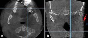
Axial cut (A) and coronal cut (B) of CBCT sialography show stenosis of the ductules with punctuated appearance of the gland (arrow).